Cellular pathologists play a vital role in developing a greater understanding of disease and abnormalities in organs, tissue and cells. As technology develops, cellular pathology is evolving to further support public health. Discover more about the key achievements of this specialty from the advent of immunohistochemistry and molecular pathology to the standardisation of cancer reporting.
As a discipline, cellular pathology represents a fascinating mixture of traditional methods and techniques, together with an ever-evolving role due to changes in other clinical specialties and the introduction of new technologies. The specialty has become increasingly important to medicine, both in initial diagnosis and guiding patient management. While this has placed increasing pressure on our service that is sometimes difficult to manage, it has also made cellular pathology an even more interesting and fulfilling specialty to be involved in. Change within cellular pathology has accelerated over the last decade and we are now embarking on a very exciting period of further developments that will result in a modernised service, fit for the evolving NHS as we move forward over the next few years.
Key achievements
Immunohistochemistry
Immunohistochemistry entered diagnostic practice in the 1980s and is now a mainstream method in cellular pathology laboratories. The technique is based on the identification of characteristics of cells and tissues via a specific, immune-based interaction between a primary antibody raised in a non-human animal and the cellular or tissue component under investigation. Immunohistochemistry has become a cornerstone of many aspects of diagnostic practice, including the typing and subtyping of tumours and the identification of infective agents. Immunohistochemistry has also become increasingly important for the prediction of clinical response to cancer-targeted therapies.
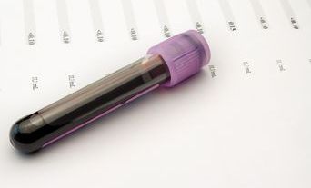
Molecular pathology
Molecular pathology describes the very broad range of techniques that are primarily based on the investigation of DNA or RNA for many purposes, mainly associated with neoplasia. Some techniques can be performed on tissue sections mounted on glass slides. However, many techniques are centred on the examination of DNA that is extracted from fresh or formalin-fixed tissues. Almost all these methods rely on the ability to amplify DNA samples using the polymerase chain reaction (PCR). Kary Mullis, the inventor of PCR, shared the Nobel Prize for Chemistry in 1993 for this discovery.
Nowadays, molecular tests are essential for the initial diagnosis of many neoplastic conditions, for instance, the identification of clonality in lymphomas and leukaemias, as well as for the determination of prognosis and the prediction of clinical response to an increasing range of cancer-targeted treatments. Molecular techniques are also used for other reasons, such as the identification of infective agents.
Cellular pathology represents a fascinating mixture of traditional methods and techniques, together with an ever-evolving role due to changes in other clinical specialties and the introduction of new technologies.
Cancer datasets and tissue pathways
The quality and complexity of cellular pathology reports has increased dramatically over the last 30 years. By the mid-1990s, it had become clear that the amount of information in reports for cancer resections was variable. At the same time, research linking cellular pathology with patient outcomes was revealing an increasing number of features identifiable within tissue biopsy and surgical resection specimens that were important for accurate pathological cancer staging and other aspects of disease prognostication. The College recognised the need for detailed guidance on cancer specimen handling and reporting and commissioned the creation of a series of publications on an anatomical site-specific basis. These were initially called Minimum datasets but were later renamed as Datasets for the histopathological reporting of cancers, with the first appearing in 1998.
Over the subsequent two decades, the range of cancer sites covered by these datasets has increased progressively, such that there are currently over 40 datasets covering all major cancers. The publications comprise a detailed and fully referenced text providing guidance on all aspects of specimen dissection and block selection, together with one or more proformas that include all the required data items. Each dataset is also regularly updated by experts in the field such that new developments in clinical management, cellular pathology and molecular pathology are included as they become relevant to diagnostic practice.
Biomedical and clinical scientists represent a key component of cellular pathology staffing. Within many areas of medicine, roles previously undertaken only, or mainly, by medical staff are now being performed by nursing or scientific staff.
Subspecialisation in larger centres
Until the 1990s, almost all cellular pathology departments were staffed by histopathologists and/or cytopathologists who were trained in all major organ systems and specimen types and whose routine diagnostic practice was across all major subspecialties, although they may well have possessed one or more areas of special interest and expertise.
During the 1990s, clinicians became increasingly subspecialised, with the days of the truly general surgeon and physician effectively over. In parallel with developments in medicine, the practice of cellular pathology was becoming more complex, which was reflected in the creation of the cancer datasets described in the previous section.
The concept of formal subspecialisation in cellular pathology developed initially in larger and usually academic departments. This would allow pathologists to focus on one, or a small number, of areas within diagnostic practice, facilitating the maintenance of up-to-date knowledge and the acquisition of specialist skills that were particularly pertinent to the most complex and/or unusual cases. This process has since become widespread within large departments and is also increasingly common within smaller hospital departments, once the number of histopathologists and/or cytopathologists exceeds the minimum required in order to make such an approach viable and sustainable.
Scientist input to complex specimen cut-up
The relentless increase in workload and specimen complexity within cellular pathology has led to increasing pressure on most departments in the UK.
Despite initiatives to increase the number of medically trained graduates entering the discipline with the creation of histopathology training schools across the UK and increases in the numbers of funded training posts, many departments have become understaffed in terms of the required number of trained individuals to perform specimen cut-up and reporting duties.
Biomedical and clinical scientists represent a key component of cellular pathology staffing. Their roles have traditionally focused on assistance with cut-up, tissue processing, the preparation of tissue sections and both histochemical and immunohistochemical staining.
Within many areas of medicine, roles previously undertaken only, or mainly, by medical staff are now being performed by nursing or scientific staff. Training scientists to perform specimen cut-up and expanding this role to more complex cases, under the guidance of the medical staff, is a significant service improvement that has also created an interesting career pathway for this staff group.
Key challenges facing cellular pathology
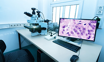
Digital pathology
The introduction of digital technology into cellular pathology is an exciting development that will bring investment into laboratories and create image-handling capabilities that will facilitate aspects of routine diagnostic work and many academic pursuits involving this discipline. Key challenges include ensuring that diagnostic accuracy is maintained during the introduction of digital systems. It will also be important that systems in different hospitals can interact and allow digital image transfer between departments. It is essential to ensure that changes in working patterns that could follow the introduction of digital pathology, such as increased working from home, are appropriately managed.
Multidisciplinary team meeting support
Multidisciplinary team meetings were introduced following the 1995 Calman–Hine report.1 The concept was to ensure that the highest possible standard of care was delivered to cancer patients through formal involvement of every key discipline involved in their management. This principle has expanded throughout cancer and non-cancer care and has been an important means of ensuring that the best care is provided. Ensuring that these meetings are appropriately resourced and that pathologist input is streamlined where possible are crucial challenges.
We are now embarking on a very exciting period of further developments that will result in a modernised service...
Molecular pathology
Molecular pathology is now a key component of modern patient care for the reasons described earlier in this article. Our main challenges include ensuring that the sometimes-complex administrative pathways involved in arranging these tests operate smoothly and that the pre- and post-test activities required of cellular pathology laboratories are recognised and appropriately funded.
Developing systems for cellular pathology
Laboratory information management systems (LIMS) across the UK are often relatively old-fashioned compared to the computer software available in other areas of modern life. Further-more, procuring a new LIMS is almost always a protracted process. The development of the Carter report-inspired hub and spoke model for the provi-sion of pathology services within the NHS will be greatly facilitated by the introduction of region-wide LIMS that allow secure data transfer between laboratories. Another challenge is ensuring that new LIMS support the latest developments within cellular pathology. For example, use of well formatted proforma-style reports, interfacing with digital pathology and robust systems for acknowledgement of results.
Facilitating funding
Cellular pathology has historically struggled to obtain funding to maintain and expand its service. There is a tendency for service users to assume that changes within their own disciplines will not affect the workload of cellular pathology – or to fail to recognise workload implications for our specialty. Key challenges include maintaining service-wide surveillance for new developments that may impact on cellular pathology and engaging with service users to ensure that these are recognised with an appropriate level of funding.
The tighter integration of our specialty with patient pathways should help to raise the profile of cellular pathology and facilitate this process.
Securing sufficient staffing
The increasing workload of cellular pathology departments (in terms of both case numbers and complexity) has not been paralleled by a sufficient increase in medical, scientific and administrative staffing across the service as a whole. Marked differences in staffing levels exist across UK departments, with the smaller laboratories often least able to recruit and retain staff. Key challenges include securing funding to enable staff expansion and then successfully recruiting to fill vacancies. The introduction of digital pathology and regional LIMS should facilitate the geographical flexibility of service support to laboratories most in need of assistance.
Integrating scientists into reporting
Biomedical and clinical scientists are a key component of cellular pathology departments. Scientist-led support of roles traditionally undertaken by medically trained pathologists can provide valuable service support. Many departments have already integrated scientists into specimen dissection. Over 50 laboratories in the NHS are now training scientists to undertake microscopy and reporting, within defined areas of practice. Incorporating scientists into these tasks in a way that is most beneficial to the service is a key challenge. It is essential to approach this development from the point of view of positively determining what scientists can achieve and contribute. It is important to recognise that this development is similar to many initiatives involving non-medically qualified staff across the whole of clinical medicine, for instance nurse-led endoscopy.
Celebrating notable cellular pathologists
Juan Rossi
Juan Rosai is seen by many as the father of modern cellular pathology. He was born in Italy but undertook his medical degree and initial pathology training in Argentina. He worked in several high-profile positions at Washington University, the University of Minnesota, Yale University and the Memorial Sloan-Kettering Cancer Center in New York. He later returned to Italy, to the Istituto Nazionale dei Tumori in Milan. As his reputation as a surgical pathologist developed, he received accolades from many institutions including the International Academy of Pathology, the American Board of Pathology and the Royal College of Pathologists. His expertise as a generalist diagnostician was unparalleled – a skill that is now unlikely to be encountered in the era of increasing subspecialisation. He published over 400 papers, many of which contained seminal descriptions of new entities, especially in thyroid, thymic and vascular pathology. He was editor-in-chief of the third series of the Atlas of Tumour Pathology of the Armed Forces Institute of Pathology and an editor of the World Health Organisation Classification of Tumours series. He is probably best known internationally for Rosai and Ackerman’sSurgical Pathology – the most famous benchmark general text within cellular pathology. He was also a renowned teacher and mentor.
Basil Morson
Basil Morson was, and remains, the doyen of gastrointestinal pathology worldwide.
He was born in London, served in the Royal Navy during the Second World War and later joined the Royal Naval Reserve. He studied for his medical degree and started his pathology training at the Middlesex Hospital Medical School in London. He gained MA and DM degrees at Oxford University. In the 1950s, he was the first to describe gastric-type metaplasia in the disease that became known as Barrett’s oesophagus and undertook innovative work on intestinal metaplasia in the stomach as a precursor of gastric cancer.
Later, he started working with Dr Cuthbert Dukes at St Mark’s Hospital, London, and was subsequently appointed as his successor in 1956. He was particularly attracted to Dr Dukes’ work on surgical specimens of colorectal cancer. Basil was very influential in the development of the endoscopic biopsy and wrote seminal articles on the pathology of such material. Despite working as a single-handed pathologist in a small specialist hospital, he undertook much innovative research. He wrote important papers on the distinction of Crohn’s disease from ulcerative colitis, authored the initial description of the biopsy appearances of dysplasia complicating ulcerative colitis and produced ground-breaking work on the concept of the adenoma-carcinoma sequence in the large intestine in the 1970s.
He was also an excellent administrator. He was Vice President and Treasurer of the Royal College of Pathologists and was also President of the British Division of the International Academy of Pathology and of the Sections of Proctology and United Services of the Royal Society of Medicine. He was the first pathologist to be the President of the British Society of Gastroenterology and his contributions were recognised by in 1987 with the establishment of the Basil Morson Lecture, still the premier named lecture in gastrointestinal pathology. He established and co-authored Morson and Dawson’s Gastrointestinal Pathology, still the flagship UK text on this subject, with the sixth edition to be published soon. He was the author of 11 other books, 20 book chapters and over 200 original papers. He greatly valued his interactions with clinicians and would often begin a lecture to trainee pathologists with the phrase, ‘it is your job to control surgeons’, intimating that one inappropriate word on a pathology report may induce unindicated major resections. In 1987, he was awarded a CBE for services to medicine.
Author(s)

Adrian Bateman

Newton Wong