
What is a biopsy?
A biopsy is a medical procedure that involves taking a small sample of tissue so that it can be examined under a microscope. A tissue sample can be taken from almost anywhere on, or in the body, including the skin, stomach, kidneys, liver and lungs. The term biopsy is often used to refer to both the act of taking the sample and the tissue sample itself.
What is a biopsy used for?
Biopsies can be used to investigate the cause of a person’s symptoms or to help diagnose a number of different health conditions. Where a condition has already been diagnosed, a biopsy can be used to measure how severe it is or at what stage it is. For example, the results of a biopsy can show how severely an organ, such as the liver, is inflamed.
It is impossible to be sure whether a lump or growth on the skin or inside the body is cancerous (malignant) or non-cancerous (benign) just by looking at it or feeling it.
A biopsy can provide this information. Ruling out a diagnosis is much more difficult and may require several tests including multiple biopsies.
Types of biopsy
There are various types of biopsy that can be used to help identify a wide range of health conditions. How a biopsy is carried out will depend on from where the tissue sample is being taken.
An operation can start with a biopsy with the sample being tested straight away so that the surgeon can carry out appropriate surgery using the diagnosis provided.
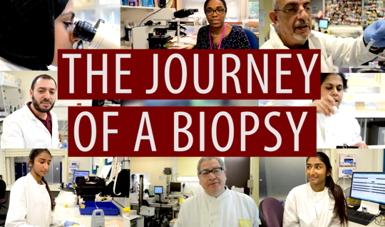
The role of the histopathologist
Histopathologists are the doctors who diagnose cancer and other serious illnesses – but they also often have good news, for example discovering that a lump or mole is completely benign. Some histopathologists also carry out autopsies (post-mortems) to find out why someone has died. Histopathologists are also at the forefront of research into many common diseases such as cancer.
Histopathologists examine biopsies (tissue or cells) removed from patients in the clinic or during an operation.
The histopathologist examines tissue biopsies with the naked eye to look for any visible abnormalities and to select pieces to examine in more detail under the microscope. These small pieces are treated with chemicals so that very thin slices can be cut. The slices are stained to show different parts of the cells and examined under a microscope to see whether the tissue is abnormal. If it is, the aim is to identify the nature of the problem. This often means that a definite diagnosis is made. They also examine cells in bodily fluids (cytopathology) such as urine, and also in large specimens, for example from surgery for bowel or breast cancer. These specimens are dissected (‘cut up and trimmed’) to select the most appropriate areas to examine under the microscope.
After the tissue sample has been removed, it can be tested using various chemicals to see how it responds and to find out what it contains. The type of tests used will depend on the medical conditions being investigated and the features of the tissue sample.
Pathologists attend multidisciplinary team (MDT ) meetings where the pathology findings are interpreted in the context of the whole of the patient’s treatment and care and discussed with other clinicians such as surgeons and oncologists. Treatment plans are then devised for each individual patient.
A MDT team meeting is a meeting of a group of professionals from several clinical disciplines who make decisions together regarding recommended treatment of individual patients. MDTs may specialise in certain conditions, such as a specific cancer.
-

Dr Rachel Brown, Histopathologist
My areas of interest are liver/pancreas and head and neck pathology so I might be looking at a liver removed at transplantation or a partial liver resection or pancreatic resection for tumour.
